Echocardiogram
What is an echocardiogram?
A transthoracic echocardiogram (TTE) is a type of test that utilises ultrasound waves to create images of the heart, allowing the doctor to check the structure and function of the organ.
When do I need to go for a echocardiogram screening?
A transthoracic echocardiogram is recommended to diagnose and monitor various heart conditions and other medical conditions. Besides identifying heart conditions, transthoracic echocardiogram can help your doctor identify and evaluate the following:
- Evaluate the function of the heart
- Determine the appropriate treatment plan
- Monitor heart health before and after diagnosis
- Determine if there are any problems with the heart, valves, or blood vessels
- Diagnose any congenital heart diseases
Are there any side effects of a echocardiogram?
There are no side effects associated with a transthoracic echocardiography. However, you may experience some discomfort during the screening as the ultrasound transducer will be held firmly against your chest. This firmness is needed to ensure the images taken of your heart and blood vessels are of high quality.
How do I prepare for a echocardiogram?
Preparing for a transthoracic echocardiogram is straightforward and requires minimal preparation. To ensure a smooth screening test, it is recommended to do the following:
- Wear comfortable and avoid tight-fitting clothes
- Avoid consuming any food or fizzy drinks 2 hours before the test
- Wear clothing that is easily removable from the waist up
- Take your medications at the regular times
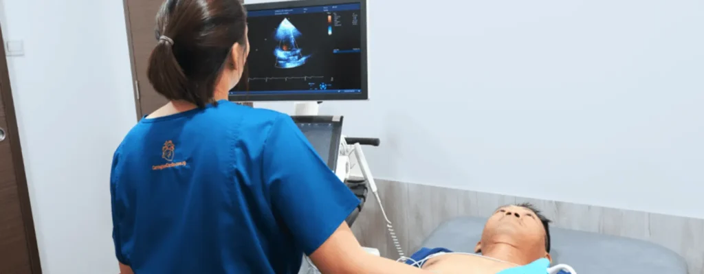
What happens during a echocardiogram?
A transthoracic echocardiogram usually takes between 30 to 60 minutes to complete, and you will be lying mostly on your left side during the test. Prior to the test, a sonographer will take the necessary height, weight, and blood pressure measurements. After taking your biometrics, you will be asked to remove any clothing from the waist up and the steps during the test are as followed:
- A sonographer will place several small, sticky patches on your chest, which are connected to an ECG machine to monitor your heart rhythm.
- Next, some ultrasound gel will be placed on your chest and upper abdomen to help improve the quality of images.
- The sonographer will use a tool (transducer) to move back and forth on the chest to take different images of your heart at different angles.
- At the end of the test, the ECG patches are removed, and the gel will be wiped away from your chest.
What should I be doing after a echocardiogram?
After the test, you can resume your normal daily activities as there is no recovery time or any specific restrictions. Once the images have been reviewed by the cardiologist, we will reach out to you to discuss your next appointment booking. During the appointment, the cardiologist will discuss the results, diagnosis and treatment plan.